Judgment Call: Exploring Treatment Tactics in Clinically Isolated Syndrome
More aggressive strategies in the treatment of multiple sclerosis have created an opportunity for physicians to refine the way they address a first demyelinating event, and perhaps change the course of disease.


Ben Thrower, MD
Recent evidence suggests that the pathological process of multiple sclerosis (MS) is already underway at the time of the first episode of clinically isolated syndrome (CIS), defined as sudden neurologic dysfunction due to inflammation and demyelination that lasts 24 hours or more and does not include signs of fever, infection, or encephalopathy.1-3 As the range of effective treatment options for MS has expanded and the ability of magnetic resonance imaging (MRI) to track changes of pathology in response to therapies has improved, the rationale for treating CIS has grown stronger.
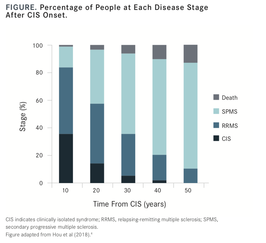
Although up to one-third of patients with CIS will have no further demyelinating events, a 2018 article exploring the natural course of CIS showed that more than 85% of patients diagnosed with CIS progressed to relapsing-remitting or secondary progressive MS within 20 years (FIGURE).4 The challenge lies with predicting which of those patients will experience future events and how to mitigate potential damage in the interim.1,2
The Rationale for Early Diagnosis
Although CIS has been shown to resolve with or without treatment, evidence accumulated over the past several years suggests that the implications of untreated CIS are significant to the later course of MS.
In the early period following CIS, much of the activity associated with MS remains subclinical, but it is still detectable on MRI. In fact, MRIs taken during this period show substantial axonal damage occurring despite the resolution of symptoms. A 2007 review by Ben W. Thrower, MD,2 suggested that typical demyelination patterns seen on traditional MRIs appeared as “islands” of axonal pathology surrounded by normal-appearing white matter. Newer developments in MRI technology, however, have revealed these islands to be more like icebergs, with an estimated 50% of untreated MS cases showing enhancing lesions.2 Nearly one-third of enhancing lesions will evolve into “black holes” that correlate directly with later disability, pointing to the need for treatment at the earliest point of detection—or CIS.
In an interview with NeurologyLive®, Thrower, the medical director of the Andrew C. Carlos Multiple Sclerosis Institute at the Shepherd Center in Atlanta, Georgia, explained that the main risk associated with early diagnosis of MS is that “you want to make sure you’ve gotten it right, because once a person has gotten an MS diagnosis, sometimes there’s not a lot of additional thought about the diagnosis. People just go with the flow and stay on a treatment that is maybe not appropriate for them, and whatever the correct diagnosis is, it’s not being managed at that point.
“We need to consider a balance in that you want to diagnose quickly, but also diagnose accurately,” Thrower continued. “At every [MS treatment] center, you’ll probably have a small handful of patients who have been misdiagnosed, because [they have] some- thing that mimics MS.”


Diagnosis of MS at First CIS
Expansion of the original 2010 McDonald Criteria, which specified dissemination of lesions in time and space, now allows for much earlier diagnosis of MS.5 The 2017 update to the McDonald Criteria states that with evidence of dissemination in space (DIS), the requirement for clinical or MRI criteria for dissemination in time (DIT) can be met by the presence of oligoclonal bands in the absence of atypical cerebrospinal fluid findings; with no better explanation, this allows for a diagnosis of MS.5 Other changes to the diagnostic criteria include the recommendation that both symptomatic and asymptomatic MRI lesions can be applied to the determina- tion of DIS and DIT, and that cortical lesions can substitute for DIS.5
“MS can be diagnosed with 1 MRI, if both an old and new lesion are detected at the time of CIS,” Thrower said. “Everything with multiple sclerosis always comes back to our original classic definition of dissemination of focal neurological events in time and space. And really, with 1 MRI, we can see that dissemination in time and space if we have 1 clinical event, and we have evidence of both old and new lesions on MRI. I think we’re just using technology to be able to meet that definition earlier.”
Optic neuritis (ON) and other manifestations of CIS have predictive value as well. The new criteria state that young female patients who present with unilateral ON that is painful have a higher risk of future demyelinating events, even with normal fundoscopy. Asymmetric presentation of transverse myelitis, when combined with a nonedematous spinal cord lesion, is also associated with a greater risk of MS.5
“We’re now using a new blood test for myelin oligodendrocyte glycoprotein to aid in the diagnosis of MS and similar conditions,” Thrower added. “Some new [research has] looked at the morphology of MRI lesions—whether they enhance, where they’re located, and how to determine risk for clinically definite MS and disease progression.”
Now that a diagnosis of MS can be made earlier, the question becomes whether it should be treated.
“Yes,” Thrower stated simply. “And the driving force behind that, if you look at our goals for treating a newly diagnosed individual, is that we would like to achieve no evidence of disease activity (NEDA). No evidence, meaning they have no relapse, no increased disability, and no MRI lesions.” He noted that setting the bar that high may not really be targeting the patient’s whole condition at the time. There may still be damage occurring that is at a level below what is visible given the resolution of standard MRI techniques, and those possibilities should be considered. “A patient on interferon may look like they are achieving NEDA, but really a level of damage is occurring beneath the surface. We may see them paying for that 10 or 15 years down the line,” he said.
Approaches to Treating CIS
Treatment approaches to CIS generally follow the same standards as those for treatment of clinically defined MS, with disease modifying therapies (DMTs) as the first-line treatment preferred by most clinicians. The growing understanding of how early demy-elinating processes may already be at work at the time of CIS has been a driving force behind more aggressive treatment strategies. It becomes important, Thrower said, to confirm the diagnosis early and to at least put more effective therapies on the table for discussion with the patient and their family as first-line options.
“We have 19 FDA-approved treatment options. They all have a certain safety profile and efficacy profile, and some drugs are more effective than others,” said Thrower. But, he explained, a para- digm shift has occurred in how existing DMTs are being utilized in MS, and especially at the time of CIS, that encompass 2 different schools of thought: escalation therapy versus induction therapy.
Escalation Therapy
The more traditional approach to MS treatment that is frequently applied to patients with CIS is escalation therapy. This involves initiating treatment in a newly diagnosed individual with one of the safer drugs. Such a drug may show only modest efficacy, and the patient is escalated to more aggressive therapies if and when treatment failure occurs.6 Glatiramer acetate and interferon beta are considered first-line therapies for this approach, with immunosup-pressants as second-line treatments.6
Induction Therapy
Alternatively, many MS providers are shifting to an induction strategy, which focuses on using the most effective therapies that can be used safely from the initial onset of CIS. Over time, numerous drugs, including mitoxantrone, natalizumab, and alemtuzumab, have been investigated for induction therapy in MS, all with success.6
Today, however, the main choices for induction therapy are natalizumab and ocrelizumab, followed by fingolimod and siponimod, Thrower said. “If you ask providers at different MS centers, you may get some variations in the answers, but I would say natalizumab, ocrelizumab, and alemtuzumab are probably our 3 most effective therapies,” noted Thrower, referring to his experience at the Shepherd Center. “For most [clinicians], the safety profile of alemtuzumab is not going to make it an acceptable first-line therapy. However, ocrelizumab and natalizumab are acceptable first-line therapies. With natalizumab, it really hinges on the JC virus (JCV) antibody index to determine whether someone is a good candidate. If someone is JCV antibody—negative or has a low index value, natalizumab may be a great first-line option.”
Use of these more aggressive therapies presents additional risks, particularly to newly diagnosed patients, but the risks are increasingly being outweighed by the benefits. Thrower believes that when appropriate intervention is delivered early enough, the underlying damage that was often historically present at the typical point of MS diagnosis can be averted so that progression doesn’t occur. “We have individuals on some of these more effective therapies who are just stable for years,” he said.
The Ongoing Role of MRI
While MRI in MS has largely been used for diagnosis and to monitor progression and treatment efficacy, novel techniques developed only recently have significantly enhanced the detec- tion of lesions and new pathology that could not be discerned on conventional MRI.2 This new degree of sensitivity vastly expands its utility in therapeutic strategy.
“We were using MRI in the initial diagnosis to confirm MS and to rule out other conditions,” Thrower said. “Again, the goal is NEDA, and one-third of NEDA is no new or active enhancing lesions on MRI. So, the average person is now going to get an MRI of the brain and spine on a yearly basis.”
MRI is also often utilized at the time of initiating a new treatment or changing to another drug to help confirm efficacy. “If you’re seeing evolution of enhancing lesions, you might switch to another therapy that avoids provocation of new lesions,” Thrower said.
More recently, MRI has been used to monitor subclinical activity in patients in the earliest stages of disease and to predict long-term outcomes. In a 2018 study,7 Céline Louapre, MD, PhD, of Institut du Cerveau et de la Moelle Epinière in Paris, France, reported that new MRI techniques “offer insights into MS pathophysiology beyond white matter lesions.” For instance, new evidence shows that emer- gence of new lesions in specific regions, such as the medullary and infratentorial areas of the brain, are highly predictive of long-term disability and higher risk of progression to secondary MS.7
Thrower pointed out that while taking more frequent MRIs may provide more information, it’s important to look at the patterns over time for context. “That once-a-year MRI may change over time. If we have a patient who is just absolutely stable over 5 or 1 years—there’s no set number—then we would potentially do MRIs less frequently, similar to progressive forms of MS where the MRIs don’t change as drastically.”
Thrower also emphasized the importance of helping the patient understand what the changes—or lack of changes—to MRIs means for their disease course and treatment. “We always let patients know, too, that MRI is a great tool, but you have to put it into the context of how the patient is doing clinically. Early in MS you can have new disease activity that’s largely asymptomatic, where they come in and the MRI shows a new lesion. Later in MS ,you can see individuals whose MRIs really aren’t changing, but they’re progressing—and that’s very frustrating for the patient. When I tell a patient who is getting worse that their MRI is stable, at some level they hear that you don’t believe them. We do a lot of education around that,” he said.
“If you look at MS management prior to 1993, when we had no treatment options, some of those treatment paradigms were based on in-patient models, and it was just assumed that every- body was going to progress eventually. Now, 99.9% of MS therapy is outpatient, and I do think we’ve changed the natural history curve dramatically. I feel reasonably good about the potential of a newly diagnosed individual—if we get them on the right treatment early— that hopefully we’ll be able to put the brakes on MS and not see progression over decades,” Thrower concluded.
REFERENCES
1. Brownlee WJ, Miller JH. Clinically isolated syndromes and the relationship to multiple sclerosis. J Clin Neurosci. 2014;21(12):2065-2071. doi: 10.1016/j.jocn.2014.02.026.
2. Thrower BW. Clinically isolated syndromes: predicting and delaying multiple sclerosis. Neurology. 2007;68(24 suppl 4):S12-S15. doi: 10.1212/01.wnl.0000277704.56189.85.
3. Kennedy P. Impact of delayed diagnosis and treatment in clinically isolated syndrome and multiple sclerosis. J Neurosci Nurs. 2013;45(6 suppl 1):S3-S13. doi: 10.1097/JNN.0000000000000021.
4. Hou Y, Jia Y, Hou J. Natural course of clinically isolated syndrome: a longitudinal analysis using a Markov model. Sci Rep. 2018;8(1):10857. doi: 10.1038/s41598-018-29206-y.
5. Thompson AJ, Banwell BL, Barkhof F, et al. Diagnosis of multiple sclerosis: 2017 revisions of
the McDonald criteria. Lancet Neurol. 2018;17(2):162-173. doi: 10.1016/S1474-4422(17)30470-2.
6. Edan G, Le Page E. Induction therapy for patients with multiple sclerosis: why? when? how? CNS Drugs. 2013;27(6):403-409. doi: 10.1007/s40263-013-0065-y.
7. Louapre C. Conventional and advanced MRI in multiple sclerosis. Rev Neurol (Paris). 2018;174(6):391-397. doi: 10.1016/j.neurol.2018.03.009.